Conditions Explained
Disclaimer:
This website is intended to assist with patient education and should not be used as a diagnostic, treatment or prescription service, forum or platform. Always consult your own healthcare practitioner for a more personalised and detailed opinion
Cholesteatoma
We have selected the following expert medical opinion based on its clarity, reliability and accuracy. Credits: Sourced from the website Patient UK, authored by Dr Oliver Starr and Dr Helen Huins (see below). Please refer to your own medical practitioner for a final perspective, assessment or evaluation.
Overview
What is a cholesteatoma?
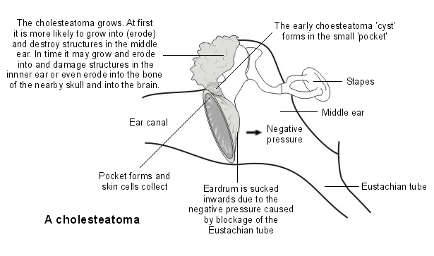
Cholesteatoma is the name given to a collection of skin cells deep in the ear that form a pearly-white greasy-looking lump deep in the ear, right up in the top of the eardrum (the tympanic membrane).
A cholesteatoma is rare. The truthful answer to what causes it, is 'we don't really know'. Skin cells from the lining of the ear canal seem to get trapped in the middle ear (which doesn't normally contain skin cells).
Skin cells, including those that line the ear canal, normally multiply regularly to replace those that have died. Usually these skin cells just flake off.
The dead cells are trapped too and build up. This build-up of dead skin cells over time is what forms the cholesteatoma.
What type of problem is a cholesteatoma?
Cholesteatoma is an uncommon condition where a cyst-like growth develops in the ear. It can be a birth defect (congenital problem) but usually occurs as a complication of long-standing (chronic) ear infection.

The most common symptoms are loss of hearing and a foul-smelling discharge from the ear. It is nota cancerous (malignant) condition but is important because it can lead to serious complications such as permanent deafness and life-threatening illnesses such as meningitis.
Symptoms
What symptoms does a cholesteatoma cause?
Symptoms start very gradually, over several months.
- The first symptom is a discharge from one ear. It is usually slightly watery, sometimes with a green or yellow colour.
- The discharge might be slightly smelly.
- This often looks to a doctor like an external ear infection (otitis externa) or an infection of the inner ear (otitis media) with a perforated eardrum.
- Because it looks just like these common infections, it is usually treated (wrongly) with antibiotic ear drops or pills.
- Although it might get slightly better with these treatments, it will never fully clear up.
- It is not painful.
- After a while you may get hearing problems in that ear.
- If the cholesteatoma is left untreated it can spread into the balance centres of the inner ear, causing dizziness.
- Eventually, in very rare cases, it can spread right next to the brain and cause an infection in the brain tissue or the lining of the brain. This is very unlikely to happen these days in the western world.
How do we hear?
The ear is divided into three parts - the external ear, the middle ear and the inner ear. The middle ear, which is behind the eardrum (the tympanic membrane) is filled with air. Air comes from the back of the nose up a thin channel called the Eustachian tube. In the middle ear there are three tiny bones (ossicles) - the hammer (malleus), anvil (incus) and stirrup (stapes). The inner ear includes the cochlea and semicircular canals.
Sound waves come into the external ear and hit the eardrum. The sound waves cause the eardrum to vibrate. The sound vibrations pass from the eardrum to the ossicles. The ossicles then transmit the vibrations to the cochlea in the inner ear. The cochlea converts the vibrations to sound signals which are sent down a nerve from the ear to the brain, allowing us to hear.
The semicircular canals in the inner ear contain a fluid that moves around as we move into different positions. The movement of the fluid is sensed by tiny hairs in the semicircular canals which send messages to the brain down the ear nerve to help maintain balance and posture.

Causes
Why does a cholesteatoma develop?
The cause of a cholesteatoma is quite difficult to explain, and even now is not fully understood.
We all have skin inside our ear canal. It is meant to be there and is a normal part of our ear. But with a cholesteatoma the skin right next to the eardrum, deep in the ear, gets sucked in gradually to where it shouldn't be. No one quite knows why this happens but it is usually related to the eardrum being very retracted (drawn inwards, deeper than it is meant to be).
This skin then forms a tiny pearl, or ball, that keeps burrowing its way deeper into the ear over many months. It damages the delicate bones inside the middle ear - the bit that is responsible for hearing. At this point it becomes painful.
If left untreated it will push further and further inside the ear, through the inner ear and possibly even next to the brain. In the western world it would be very unusual for it to get that bad but this can happen in the developing world.
There are two types of cholesteatoma:
- Congenital cholesteatoma: is a problem that in theory happens from birth. For some reason, even though the eardrum is normal, tiny skin cells get sucked into the middle ear, blocking the Eustachian tube. This then causes long-term fluid in the middle ear (which is usually free of fluid) and can cause hearing loss. This becomes apparent between the ages of 6 months to 5 years when the child's hearing doesn't develop properly. This is a very rare condition and the cause isn't fully known.
- Acquired cholesteatoma: develops later, usually in adults between 30 and 50 years old. Again, the cause isn't fully known. Sometimes a cholesteatoma in an adult can happen from having a grommet - a tiny tube that is put through the eardrum as a treatment for middle ear problems - as a child.
The true occurrence rate of cholesteatoma is not known. About 1 in 1,000 people with ear problems referred to ENT clinics have cholesteatoma. It has also been suggested that there is about 1 case per 10,000 population. Most cases are of the acquired type.
Diagnosis
How is a cholesteatoma diagnosed?
The GP or ear specialist (ENT doctor) may suspect cholesteatoma based on the typical symptoms. When the ear is examined with a torch (an otoscope), the cholesteatoma may be seen. Often there is a hole (perforation) in the eardrum (the tympanic membrane) too.
Because the symptoms come on slowly and mimic common ear infections, the diagnosis is often delayed.
- It is very difficult for a GP to see a cholesteatoma because usually it causes a lot of pus in the ear which blocks the view to the eardrum.
- For this reason the diagnosis is made by an ear specialist at a hospital.
- The ear specialist will use a tiny suction tube to suck away the discharge and look at the eardrum in detail with a microscope that magnifies the view.
- By looking in detail, close up, at the eardrum, a specialist can see the cholesteatoma pushing through the eardrum.
- To then see how far it has spread inside the ear, a specialist scan is needed: this is usually a CT scan (which takes about 30 seconds) or an MRI scan (which can take about half an hour).
What does a cholesteatoma look like?
In the image below, the white arrow on the left shows a cholesteatoma. The white tube underneath is a grommet that was put in to try to help the middle ear. The picture on the right shows what the surgeon has removed: a pale, greasy mass of cholesteatoma. It is just a few millimetres in size.

Do I need any further tests?
Hearing tests (audiometry) may show deafness or hearing loss and are usually performed in a hospital clinic. Samples (swabs) of the ear discharge may also be taken. The discharge often contains a germ (bacterium) called Pseudomonaswhich is responsible for the smell. A CT scan might be needed to see the extent of the damage caused by the cholesteatoma, and to plan further treatment.
Treatment
What is the treatment for cholesteatoma?
A cholesteatoma does not have a blood supply so taking antibiotics by mouth will not work at all because the antibiotics can't get into it. Antibiotic ear drops can clear away any infection around the cholesteatoma but will not treat the actual problem. Many people will have had antibiotic ear drops prescribed to them without success, before they are diagnosed with a cholesteatoma.
The treatment is done by an ear specialist (an ENT doctor) and usually consists of an operation under a general anaesthetic. The aim of the surgery is to remove the tiny balls of cholesteatoma and then to clear out part of the middle ear so air can circulate around better. This will hopefully stop the cholesteatoma coming back.
There are different types of surgery that can be done and a specialist ear doctor will advise you which operation is best. The most common is called a 'combined approach tympanoplasty' where the damaged part of the eardrum is removed and the bone at the back of the ear, the mastoid, is cleared out.
If the patient is not fit for surgery (for example, if they are very old or frail or have other serious medical conditions) then regular visits to an ear specialist will be recommended. This is so the specialist can suction out any tiny bits of wax or debris deep in the ear that can contribute to a cholesteatoma. This will not solve the problem but can keep it from getting worse.
What are the possible complications and why is treating it important?
Untreated, a cholesteatoma will slowly grow and expand. As it grows it can eat into (erode) and destroy anything in its path.
Therefore, possible complications that may develop over time include:
- Damage and eventual destruction of the tiny bones of the ear (the ossicles): If these are damaged, permanent deafness can occur.
- Damage to the mastoid bone: This is the thick bony lump you can feel behind the ear. The mastoid bone is normally filled with pockets of air (a bit like a honeycomb). Cholesteatoma can grow into the mastoid bone, causing infection and destroying it.
- Damage to the cochlea and other structures in the inner ear: This can cause permanent deafness on that side, and/or dizziness and balance problems.
- Damage to nearby nerves travelling to the face: This can cause weakness (paralysis) of some of the facial muscles.
- Cholesteatoma is often infected and this infection can spread to nearby body parts: In rare cases a cholesteatoma can erode through the skull next to the ear and into the brain. As a result of spreading infection, conditions such as meningitis and brain abscess can develop. These conditions can cause death.
Please note: although cholesteatoma sounds nasty, it is not cancerous (malignant) and does not spread to distant parts of the body.
Is any follow-up required?
Unfortunately, if you have had a cholesteatoma, you will need to be followed up for life in an ENT clinic.
You will also need to have your ears cleaned regularly at the clinic to remove wax and any dirt that has accumulated. The specialist will need to ensure that the cholesteatoma has not returned.
If the ear starts discharging again, further surgery may be required. MRI scans are increasingly being used to replace the need for further check-up surgery.
Prognosis
What is the outlook?
This depends on how much damage has been caused by the cholesteatoma by the time it is found and treated. It is also affected by whether any complications such as meningitis or deafness have occurred. The earlier surgery is done, and attending for regular follow-up, the better the chance of a good outcome.
About the authors
Dr Oliver Starr
MBChB, BMedSc, MRCS, MRCGP, DRCOG
Dr Oliver Starr is a general practitioner in Hertfordshire. He is an undergraduate tutor at University College Medical School, a general practice appraiser and a case assessor at the National Perinatal Epidemiology Unit, University of Oxford. Other interests include medical law, particularly regarding clinical negligence. Dr Starr is a council member of the Cameron Fund charity.
Dr Helen Huins
MB BS Lond, DCH, DRCOG, MRCGP, JCPTGP, DFFP
Helen qualified at Guy’s Hospital in 1989 and left London in 1990 to settle in the countryside. She works as a GP partner in a rural dispensing practice and is passionate about family medicine and continuity of care with interests in sport and nutrition. Helen has been a member of the EMIS authoring team since 1995.
_______________________________________________________________________________________________________________________
Are you a healthcare practitioner who enjoys patient education, interaction and communication?
If so, we invite you to criticise, contribute to or help improve our content. We find that many practicing doctors who regularly communicate with patients develop novel and often highly effective ways to convey complex medical information in a simplified, accurate and compassionate manner.
MedSquirrel is a shared knowledge, collective intelligence digital platform developed to share medical expertise between doctors and patients. We support collaboration, as opposed to competition, between all members of the healthcare profession and are striving towards the provision of peer reviewed, accurate and simplified medical information to patients. Please share your unique communication style, experience and insights with a wider audience of patients, as well as your colleagues, by contributing to our digital platform.
Your contribution will be credited to you and your name, practice and field of interest will be made visible to the world. (Contact us via the orange feed-back button on the right).