Back Conditions Explained
Adult Spondylolisthesis in the Low Back
We have selected the following expert medical opinion based on its clarity, reliability and accuracy. Credits: Sourced from the website OrthoInfo. Please refer to your own medical practitioner for a final perspective, assessment or evaluation.
What is adult spondylolisthesis in the low back
In spondylolisthesis, one of the bones in your spine — called a vertebra — slips forward and out of place. This may occur anywhere along the spine, but is most common in the lower back (lumbar spine). In some people, this causes no symptoms at all. Others may have back and leg pain that ranges from mild to severe.
Understanding how your spine works can help you better understand spondylolisthesis.
Types of Spondylolisthesis
Many types of spondylolisthesis can affect adults. The two most common types are degenerative and spondylolytic. There are other less common types of spondylolisthesis, such as slippage caused by a recent, severe fracture or a tumor.
Degenerative Spondylolisthesis
As we age, general wear and tear causes changes in the spine. Intervertebral disks begin to dry out and weaken. They lose height, become stiff, and begin to bulge. This disk degeneration is the start to both arthritis and degenerative spondylolisthesis (DS).
As arthritis develops, it weakens the joints and ligaments that hold your vertebrae in the proper position. The ligament along the back of your spine (ligamentum flavum) may begin to buckle. One of the vertebrae on either side of a worn, flattened disk can loosen and move forward over the vertebra below it.
This slippage can narrow the spinal canal and put pressure on the spinal cord. This narrowing of the spinal canal is called spinal stenosis and is a common problem in patients with DS.
Women are more likely than men to have DS, and it is more common in patients who are older than 50. A higher incidence has been noted in the African-American population.
Degenerative spondylolisthesis:

In this x-ray taken from the side, vertebrae in the low back have slipped out of place due to degenerative spondylolisthesis:

Spondylolytic Spondylolisthesis
One of the bones in your lower back can break and this can cause a vertebra to slip forward. The break most often occurs in the area of your lumbar spine called the pars interarticularis.
In most cases of spondylolytic spondylolisthesis, the pars fracture occurs during adolescence and goes unnoticed until adulthood. The normal disk degeneration that occurs in adulthood can then stress the pars fracture and cause the vertebra to slip forward. This type of spondylolisthesis is most often seen in middle-aged men.

(Left) In spondylolysis, a fracture often occurs at the pars interarticularis. (Right) Because of the pars fracture, only the front part of the bone slips forward.
Because a pars fracture causes the front (vertebra) and back (lamina) parts of the spinal bone to disconnect, only the front part slips forward. This means that narrowing of the spinal canal is less likely than in other kinds of spondylolisthesis, such as DS in which the entire spinal bone slips forward.
This x-ray taken from the side shows a pars fracture (arrow) and the resulting spondylolisthesis:

Symptoms
About 4% to 6% of the U.S. population has spondylolysis and spondylolisthesis. Most of these people live with the condition for many years without any pain or other symptoms.
Symptoms of Degenerative Spondylolisthesis
Patients with DS often visit the doctor's office once the slippage has begun to put pressure on the spinal nerves. Although the doctor may find arthritis in the spine, the symptoms of DS are typically the same as symptoms of spinal stenosis. For example, DS patients often develop leg and/or lower back pain. The most common symptoms in the legs include a feeling of vague weakness associated with prolonged standing or walking.
Leg symptoms may be accompanied by numbness, tingling, and/or pain that is often affected by posture. Forward bending or sitting often relieves the symptoms because it opens up space in the spinal canal. Standing or walking often increases symptoms.
Symptoms of Spondylolytic Spondylolisthesis
Most patients with spondylolytic spondylolisthesis do not have pain and are often surprised to find they have the slippage when they see it in x-rays. They typically visit a doctor with low back pain related to activities. The back pain is sometimes accompanied by leg pain.
Doctor Examination
Doctors diagnose both DS and spondylolytic spondylolisthesis using the same examination tools.
Medical History and Physical Examination
After discussing your symptoms and medical history, your doctor will examine your back. This will include looking at your back and pushing on different areas to see if it hurts. Your doctor may have you bend forward, backward, and side-to-side to look for limitations or pain.
Imaging Tests
Other tests which may help your doctor confirm your diagnosis include:
- X-rays. These tests visualize bones and will show whether a lumbar vertebra has slipped forward. X-rays will show aging changes, like loss of disk height or bone spurs.
X-rays taken while you lean forward and backward are called flexion-extension images. They can show instability or too much movement in your spine. - Magnetic resonance imaging (MRI). This study can create better images of soft tissues, such as muscles, disks, nerves, and the spinal cord. It can show more detail of the slippage and whether any of the nerves are pinched.
- Computed tomography (CT). These scans are more detailed than x-rays and can create cross-section images of your spine.
Treatment
Nonsurgical Treatment
Although nonsurgical treatments will not repair the slippage, many patients report that these methods do help relieve symptoms.
Physical therapy and exercise: Specific exercises can strengthen and stretch your lower back and abdominal muscles.
Medication: Analgesics and non-steroidal anti-inflammatory medicines may relieve pain.
Steroid injections: Cortisone is a powerful anti-inflammatory. Cortisone injections around the nerves or in the "epidural space" can decrease swelling, as well as pain. It is not recommended to receive these, however, more than three times per year. These injections are more likely to decrease pain and numbness, but not weakness of the legs.
Surgical Treatment
Surgical candidates with DS: Surgery for degenerative spondylolisthesis is generally reserved for the patient who does not improve after a trial of nonsurgical treatment for at least 3 to 6 months.
In making a decision about surgery, your doctor will also take into account the extent of arthritis in your spine, as well as whether your spine has excessive movement.
DS patients who are candidates for surgery often are unable to walk or stand, and have a poor quality of life due to the pain and weakness.
Surgical candidates with spondylolytic spondylolisthesis: Patients with symptoms that have not responded to nonsurgical treatment for at least 6 to12 months may be candidates for surgery.
If the slippage is getting worse or the patient has progressive neurologic symptoms, such as weakness, numbness, or falling, and/or symptoms of cauda equina syndrome, surgery may help.
Surgical procedures: Surgery for both DS and spondylolytic spondylolisthesis includes removing the pressure from the nerves and spinal fusion.
Removing the pressure involves opening up the spinal canal. This procedure is called a laminectomy.
Spinal fusion is essentially a "welding" process. The basic idea is to fuse together the painful vertebrae so that they heal into a single, solid bone.
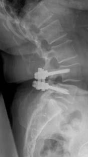
In spinal fusion, screws are often used to help stabilize the spine:

Surgical recovery: The fusion process takes time. It may be several months before the bone is solid, although your comfort level will often improve much faster.
_______________________________________________________________________________________________________________________
Are you a healthcare practitioner who enjoys patient education, interaction and communication?
If so, we invite you to criticise, contribute to or help improve our content. We find that many practicing doctors who regularly communicate with patients develop novel and often highly effective ways to convey complex medical information in a simplified, accurate and compassionate manner.
MedSquirrel is a shared knowledge, collective intelligence digital platform developed to share medical expertise between doctors and patients. We support collaboration, as opposed to competition, between all members of the healthcare profession and are striving towards the provision of peer reviewed, accurate and simplified medical information to patients. Please share your unique communication style, experience and insights with a wider audience of patients, as well as your colleagues, by contributing to our digital platform.
Your contribution will be credited to you and your name, practice and field of interest will be made visible to the world. (Contact us via the orange feed-back button on the right).
Disclaimer:
MedSquirrel is a shared knowledge, collective intelligence digital platform developed to share medical knowledge between doctors and patients. If you are a healthcare practitioner, we invite you to criticise, contribute or help improve our content. We support collaboration among all members of the healthcare profession since we strive for the provision of world-class, peer-reviewed, accurate and transparent medical information.
MedSquirrel should not be used for diagnosis, treatment or prescription. Always refer any questions about diagnosis, treatment or prescription to your Doctor.
