Back Conditions Explained
Bone Tumour
We have selected the following expert medical opinion based on its clarity, reliability and accuracy. Credits: Sourced from the website OrthoInfo. Please refer to your own medical practitioner for a final perspective, assessment or evaluation.
What are bone tumours?
Bone tumours develop when cells within a bone divide uncontrollably, forming a lump or mass of abnormal tissue.
Most bone tumours are not cancerous (benign). Benign tumors are usually not life-threatening and, in most cases, will not spread to other parts of the body. Depending upon the type of tumour, treatment options are wide-ranging—from simple observation to surgery to remove the tumour.
Some bone tumours are cancerous (malignant). Malignant bone tumours can metastasize—or cause cancer cells to spread throughout the body. In almost all cases, treatment for malignant tumours involves a combination of chemotherapy, radiation, and surgery.
Description
Bone tumours can affect any bone in the body and develop in any part of the bone—from the surface to the center of the bone, called the bone marrow. A growing bone tumour—even a benign tumour—destroys healthy tissue and weakens bone, making it more vulnerable to fracture.
When a bone tumour is cancerous, it is either a primary bone cancer or a secondary bone cancer. A primary bone cancer actually begins in bone—while a secondary bone cancer begins somewhere else in the body and then metastasizes or spreads to bone. Secondary bone cancer is also called metastatic bone disease.
Types of cancer that begin elsewhere and commonly spread to bone include:
- Breast
- Lung
- Thyroid
- Renal
- Prostate
Primary Bone Cancer
The four most common types of primary bone cancer are:
Multiple myeloma
Multiple myeloma is the most common primary bone cancer. It is a malignant tumour of bone marrow—the soft tissue in the center of many bones that produces blood cells. Any bone can be affected by this cancer.
Multiple myeloma affects approximately six people per 100,000 each year. According to the National Cancer Institute, almost 90,000 people are living with the disease each year. Most cases are seen in patients between the ages of 50 and 70. Multiple myeloma is typically treated with chemotherapy, radiation therapy and, occasionally, surgery.
Osteosarcoma
Osteosarcoma is the second most common primary bone cancer. It occurs in two to five people per million each year, with most cases in teenagers and children. Most tumours develop around the knee in either the femur (thigh) or tibia (shinbone). Other common locations include the hip and shoulder. Osteosarcoma is typically treated with chemotherapy and surgery.
Ewing's sarcoma
Ewing's sarcoma usually occurs in patients between the ages of 5 and 20. The most common locations affected are the upper and lower leg, pelvis, upper arm, and ribs. Ewing's sarcoma is typically treated with chemotherapy and either surgery or radiation therapy.
Chondrosarcoma
Chondrosarcoma is a malignant tumour composed of cartilage-producing cells. It is most often seen in patients between the ages of 40 and 70. Most cases occur around the hip, pelvis, or shoulder area. In most cases, surgery is the only treatment used for chondrosarcoma.
Benign Bone Tumours
There are many types of benign bone tumours, as well as some diseases and conditions that resemble bone tumours. Although these conditions are not truly bone tumours, in many cases they require the same treatment.
Some common types of benign bone tumours—and conditions that are commonly grouped with tumours—include:
- Non-ossifying fibroma
- Unicameral (simple) bone cyst
- Osteochondroma
- Giant cell tumour
- Enchondroma
- Fibrous dysplasia
- Chondroblastoma
- Aneurysmal bone cyst
- Osteoid osteoma
Cause
For most bone tumours the cause is unknown.
Symptoms
Patients with a bone tumour will often experience pain in the area of the tumor. The pain is generally described as dull and aching, may worsen at night, and may sometimes increase with activity.
Other symptoms of a bone tumour can include fever and night sweats.
Many patients will not have any symptoms, but will note a painless mass instead.
Although bone tumours are not caused by trauma, an injury can sometimes cause a tumour to start hurting. Injury can also cause a bone that is weakened by a tumour to fracture, or break. This may be severely painful.
Occasionally, benign tumours may be discovered incidentally when an x-ray is taken for another reason, such as a sprained ankle or knee injury.
Doctor Examination
Infections, stress fractures, and other non-tumour conditions can all closely resemble tumours. To be sure you have a bone tumour, your doctor will conduct a thorough evaluation and order a number of tests.
Medical History
As part of the examination, your doctor will take a complete medical history. He or she will ask about your general health, medications you take, and your current symptoms. Your doctor will also want to know if you or any family member has a history of any tumour or cancer.
Physical Examination
Your doctor will perform a thorough physical examination, focusing on the tumour mass.
He or she will look for:
- Swelling or tenderness in the area of the tumour
- Changes in the overlying skin
- The presence of a mass
- Any effect the tumour might have on nearby joints
In some cases, your doctor will examine other parts of your body to rule out cancers that can spread to bone.
Tests
X-rays
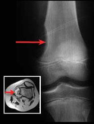
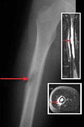
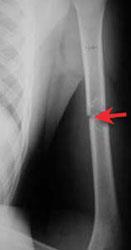
X-rays provide images of dense structures such as bone. In most cases, your doctor will order an x-ray to help diagnose a bone tumour. Different types of tumours may look different on x-ray. Some dissolve bone or make a hole in the bone. Others cause additional bone to form. Some may do both.
Femur (thighbone) tumour. This x-ray shows a tumour that caused a saucer-like erosion in the end of the thighbone. The insert shows the same tumour using a cross-sectional magnetic resonance image (MRI).
Other imaging studies
If necessary, your doctor will order a magnetic resonance imaging (MRI), computed tomography (CT), or bone scan to help further evaluate your tumour.
Femur (thighbone) tumour. This x-ray shows a tumour in the middle of the thighbone. The tumour is also seen using magnetic resonance imaging (MRI). The insert at top shows a coronal MRI. The insert at bottom shows a cross-sectional MRI. The arrows on all images show the location of the tumour.
Humerus (upper arm) tumour and fracture. This x-ray shows a fracture through a tumour in the middle of the bone of the upper arm.
Biopsy
A biopsy may be necessary to confirm the diagnosis of a bone tumour. In a biopsy, a sample of tissue is taken from the tumour. This sample is examined under a microscope and analyzed by a pathologist (a doctor who identifies diseases by studying abnormal cells).
There are two basic methods of performing a biopsy:
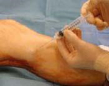
- Needle biopsy: In this procedure, you will first be given a local anesthetic. Your doctor will then insert a needle into the tumor to remove some tissue. A needle biopsy is often done in the doctor's office. In some cases, a radiologist will perform a needle biopsy. In this case, an imaging study, such as an x-ray, CT scan, or MRI scan, will be used to help direct the needle.
In a needle biopsy, the doctor inserts a needle into the tumour to remove some tissue:
.
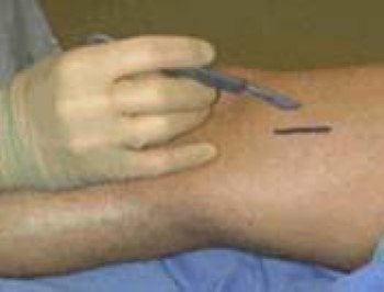
- Open biopsy: In this procedure, the biopsy is performed in an operating room. After you are given general anesthesia to put you to sleep, your doctor will make a small incision and remove some tissue.
In an open biopsy, the doctor surgically removes tissue. This is usually done in an operating room:
Other tests
Your doctor may order blood and/or urine tests to help confirm the diagnosis of a bone tumour or exclude other conditions.
Nonsurgical Treatment
Benign Tumours
If your tumour is benign, your doctor may recommend just monitoring it closely to see if it changes. During this time, you may need periodic follow-up x-rays or other tests.
Some benign tumours can be treated effectively with medication. Some will disappear over time. This is particularly true for certain benign tumours that occur in children, such as osteoid osteoma.
Malignant Tumours
If you have bone cancer, treatment will include a team of doctors from different medical specialties working together to provide care. Some will be oncologists—doctors who specialize in cancer treatment. Your team may include an orthopaedic oncologist, medical oncologist, radiation oncologist, radiologist, and pathologist. The goal of treatment is to cure the cancer while maintaining function, as best as possible, in the part of the body affected by the tumour.
Treatment depends upon several factors, including the stage of the cancer. If the cancer is localized, cancer cells are contained to the tumour and the immediate surrounding area. When the cancer has reached a metastatic stage, it has spread elsewhere in the body and may be more serious and harder to cure.
Doctors often combine several methods to treat malignant bone tumours:
- Radiation therapy: Radiation therapy uses high-dose x-rays to kill cancer cells and shrink tumours. This only treats the cancer in the area of the beam. It does not treat cancer elsewhere in the body.
- Chemotherapy (systemic treatment): Chemotherapy is often used to kill tumour cells when they have spread into the bloodstream but cannot yet be detected on tests and scans. It is generally used when cancerous tumours have a very high chance of spreading. Chemotherapy is usually given intravenously (injection into a vein) or in a pill or capsule that is swallowed.
Generally, malignant tumours are removed by surgery. Often, radiation therapy and chemotherapy are used in combination with surgery.
Surgical Treatment
Benign Tumours
In some cases, your doctor may recommend removing the tumour (excision) or another surgical technique to reduce the risk of fracture and disability. Some tumours may come back, even repeatedly, after appropriate treatment. Rarely, certain benign tumours can spread or become cancerous (metastasize).
Malignant Tumours
- Limb salvage surgery: This surgery removes the cancerous section of bone but keeps nearby muscles, tendons, nerves, and blood vessels intact where possible. The surgeon will take out the tumour and a portion of healthy tissue around it. The excised bone is replaced with a metallic implant (prosthesis), bone from elsewhere in your body, or bone from a donor.
- Amputation: Amputation is surgery to remove all or part of an arm or leg. It is usually used when a tumour is large and/or nerves and blood vessels are involved. A prosthetic limb can aid function after amputation.
Recovery
The length and complexity of your recovery will depend upon the type of tumour as well as what type of procedure was performed.
When treatment is finished, your doctor may order more x-rays and other imaging studies to confirm that the tumour is actually gone.
After treatment, you will continue to see your doctor for regular follow-up visits and tests every few months. Even though the tumour has disappeared, it is important to monitor your body for signs of recurrence. Tumours that come back may pose serious problems so it is important to detect them early.
_______________________________________________________________________________________________________________________
Are you a healthcare practitioner who enjoys patient education, interaction and communication?
If so, we invite you to criticise, contribute to or help improve our content. We find that many practicing doctors who regularly communicate with patients develop novel and often highly effective ways to convey complex medical information in a simplified, accurate and compassionate manner.
MedSquirrel is a shared knowledge, collective intelligence digital platform developed to share medical expertise between doctors and patients. We support collaboration, as opposed to competition, between all members of the healthcare profession and are striving towards the provision of peer reviewed, accurate and simplified medical information to patients. Please share your unique communication style, experience and insights with a wider audience of patients, as well as your colleagues, by contributing to our digital platform.
Your contribution will be credited to you and your name, practice and field of interest will be made visible to the world. (Contact us via the orange feed-back button on the right).
Disclaimer:
MedSquirrel is a shared knowledge, collective intelligence digital platform developed to share medical knowledge between doctors and patients. If you are a healthcare practitioner, we invite you to criticise, contribute or help improve our content. We support collaboration among all members of the healthcare profession since we strive for the provision of world-class, peer-reviewed, accurate and transparent medical information.
MedSquirrel should not be used for diagnosis, treatment or prescription. Always refer any questions about diagnosis, treatment or prescription to your Doctor.
